SPINA BIFIDA - A NEURAL TUBE DEFECT
Spina Bifida is a Latin term meaning split spine. It is the name given to a group of birth defects which interfere with the development of the central nervous system: the brain, the spinal cord and the nerve tissues.
HOW IT STARTS
The central nervous system begins to develop from the ectoderm (the layer of cells from which the brain and spinal cord develop) in the third week following fertilisation when the embryo is only 3-5mm long. Prior to this the ectoderm resembles a flat group of cells running down the middle of the embryo. This flat sheet begins to change, however, and folds to form a groove. (Diagrams (a) and (b).) The edges of the sheet eventually come together to form a tube which later develops into the spinal cord and the brain. (Diagrams (c) and (d)) This structure is called the neural tube. Once the neural tube closes it sinks into the embryo and is covered by a layer of skin. The spinal vertebrae (bony covering) then begin to form around the tube.
Normally the neural tube closes by the twenty-eighth day after conception. However, if the tube fails to close properly, a neural tube defect will occur. Neural tube defects include the conditions of anencephaly, encephalocele, and spina bifida.
SPINA BIFIDA
At some point along the spine the posterior part of the vertebrae (the bones of the spine) are not completely joined. Babies are born with the spinal cord and covering (meninges) protruding through the opening.
Within a few days of birth, the site of the lesion on the back is operated on to ensure that it has a good skin covering. This is performed to stop infection and also for cosmetic reasons.
TYPES OF SPINA BIFIDA
Possible
Spinal cord (close up) location of spina bifida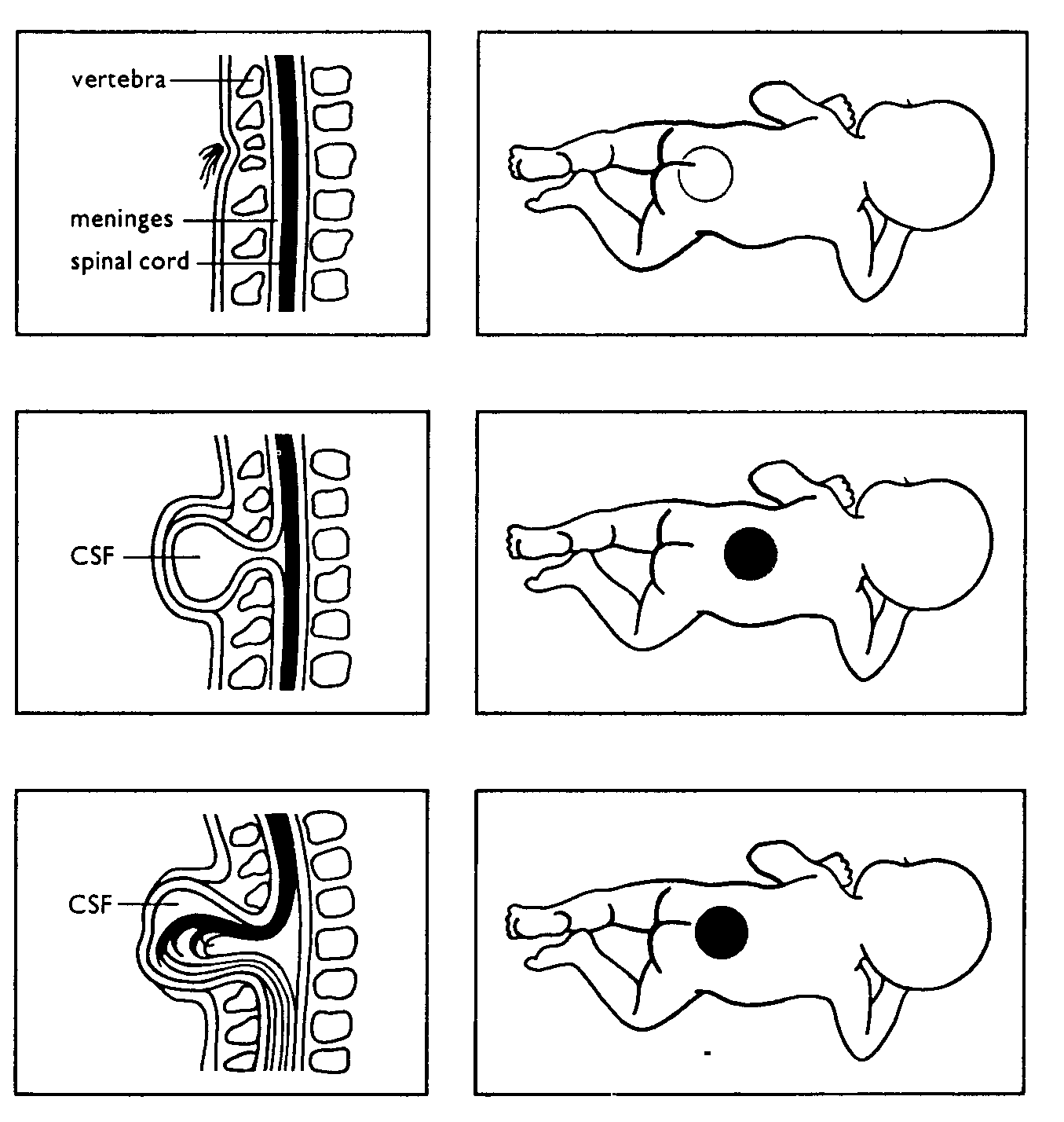
Occulta
Outer part of vertebrae not completely joined. Spinal cord and covering (meninges) usually undamaged. Hair often at site of defect.
Meningocele
Outer part of vertebrae split. Spinal cord usually normal. Meninges damaged and displaced through opening.
Myelomeningocele
Outer part of vertebrae split. Spinal cord and meninges damaged and displaced through opening. Usually hydrocephalus.
CSF Cerebrospinal fluid
Diagram reproduced from Spina Bifida and You, published by the Association for People with Spina Bifida and Hydrocephalus, 1985.
Spina Bifida Occulta
Spina bifida occulta literally means a hidden split in the spine. It is hidden because the deformity of the spine and any associated abnormalities are covered by skin. This is the least serious but most common type. For more information on spina bifida occulta.
Meningocele
In this type of spina bifida, the meninges (covering of the spinal cord) protrude through the opening, causing a lump or sac on the back. The spinal cord is often undamaged. There are usually no long-term problems, although once again, problems can arise. This is the least common form of spina bifida.
Myelomeningocele (or Meningomyelocele)
This is the most common form of spina bifida and also the most severe. The sac that has protruded on the back contains cerebrospinal fluid, blood vessels and the damaged spinal cord and meninges. There is almost always some degree of paralysis. Hydrocephalus usually occurs also.
Spina bifida most often occurs in the small of the back or lower down, but all three types can occur anywhere along the spine.
THE EFFECTS OF SPINA BIFIDA
The effect spina bifida (myelomeningocele) has on a person's life depends on many things including the location and size of the lesion, and the degree of damage to the spinal cord and nerves.
The most common occurrence of spina bifida is in the lumbar and sacral areas. The lumbar nerves control the muscles in the hip, leg, knee and foot, and help to keep the body erect. The sacral nerves control some of the muscles in the feet, bowel and bladder and the ability to have an erection. Some degree of impairment can be expected in these areas. Problems may include lack of sensation and muscle function in the lower body and legs, an inability to control urination and bowel function, joint abnormalities and deformities of the back.
WHAT ARE THE CHANCES THAT MY CHILD WITH SPINA BIFIDA WILL WALK?
This depends on many factors. However, the higher up the defect is on the spine, the more paralysis the child will probably have. The drawings below show how likely it is for the child to walk based on the level of the defect. The shaded areas show the parts of the body affected by paralysis and loss of feeling.
BONES OF THE SPINE LEVEL OF DEFECT PROBABLE AMOUNT OF DISABILITY
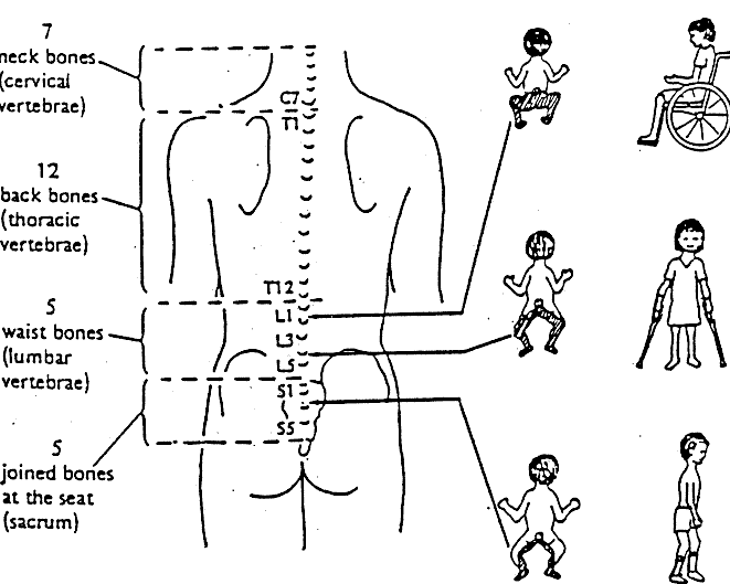 probably will not walk except with supportive equipment
* cannot control bladder or bowel
may walk using aids
later may need wheelchair
*cannot control bladder or bowel
may walk with little or no help *may or may not be able to control bladder or bowel
EXPLANATION OF SOME COMMON TERMS
ENCEPHALOCELE
Local herniation of neural tissue through a defect in the skull. Often, this will involve only the brain coverings, and children will have few problems. Sometimes, the brain is also involved. Hydrocephalus may also occur.
SCOLIOSIS
A lateral curvature of the spine, often in an S shape.
KYPHOSIS
An exaggerated outward curvature of the spine.
KYPHOSCOLIOSIS
A spinal deformity, in which both kyphosis and
scoliosis are present.
CT SCAN
An x-ray machine which revolves around the body, and takes a picture of the organs inside the body.
MRI
Magnetic Resonance Imagery - a scanner using magnetic energy to give a clear black and white picture of the brain, and spinal cord. It does not use radiation.