| Facts About Spina Bifida and Hydrocephalus |

SYMPTOMS OF A MALFUNCTIONING SHUNT
HEADACHE
VOMITING
FEVER
IRRITABILITY AND PERSONALITY CHANGES
DETERIORATION IN PERFORMANCE
school work, gait, balance, concentration
LETHARGY AND DROWSINESS
DIZZINESS AND in more severe cases VISION DISTURBANCES SEIZURES
SHUNT SYSTEM TO THE ABDOMINAL CAVITY
If any of these symptoms are present, medical advice is needed.
|
SPINA BIFIDA - A NEURAL TUBE DEFECT |
Spina Bifida is a Latin term meaning split spine. It is the name given to a group of birth defects which interfere with the development of the central nervous system; the brain, the spinal cord and the nerve tissues. |
|||||
|
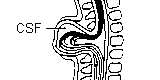
The central nervous system begins to develop from the ectoderm (the layer of cells from which the brain and spinal cord develop) in the third week after fertilisation when the embryo is only 15mm long. Prior to this period the ectoderm resembles a flat group of cells running down the middle of the embryo. This flat sheet begins to change, however, and folds to form a groove. (Diagram (a) and (b).) The edges of the sheet eventually come together and form a tube which will develop into the spinal cord and the brain. (Diagram (c) and (d).) This structure is called the neural tube. Once the neural tube closes it sinks into the embryo and is covered by a layer of skin. The spinal vertebrae (bony covering) then begin to form around the tube.
Normally the closure of the neural tube occurs around the thirtieth day after fertilisation. However, if something interferes and the tube fails to close properly, a neural tube defect will occur. Neural tube defects include, the conditions of anencephaly, encephalocele, and spina bifida. |
||||||
| In spina bifida, at some point along the spine the outer part of the vertebrae (the bones of the spine) are not completely joined. In more severe forms of spina bifida, the spinal cord and covering (meninges) protrude through the opening. Within a few days of birth, the site of the lesion on the back is operated on to ensure that it has a good skin covering. This is performed to stop infection and also for cosmetic reasons. |
||||||
|
SPINA BIFIDA OCCULTA
Outer part of vertebrae not completely joined. Spinal cord and covering (meninges) undamaged. Hair often at site of defect. |
|
|||||
|
Spina Bifida Occulta literally means a hidden split in the spine; hidden because the deformity of the spine and any associated abnormalities are covered by skin. This is the least serious type. The split in the vertebrae is so small that the spinal cord does not protrude, and so little or no damage is done. The skin at the site of the lesion may be normal, or it may have some hairs growing from it; there may be a dimple in the skin, or a birthmark. Someone with spina bifida occulta may not have any problems at all, and probably wouldn't know they had this unless an x-ray of the back was taken. Occasionally problems do arise, however, and medical advice is needed.
|
||||||
|
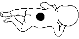
MENINGOCELE
Outer part of vertebrae Split. Spinal cord Normal. Meninges damaged and pushed through opening. |
|
|||||
| In this type of spina bifida, the meninges (covering of the spinal cord) protrude through the opening, causing a lump or sac on the back. The spinal cord is undamaged. There are usually no long-term problems, although once again, problems can arise. This is the least common form of spina bifida. |
||||||
|
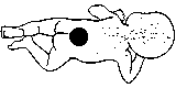
MYELOMENINGOCELE
Outer part of vertebrae not completely joined. Spinal cord and covering (meninges) undamaged. Hair often at site of defect. |
|
|||||
|
This is the most common form of spina bifida and also the most severe. In this case, the sac that has protruded on the back contains fluid, blood vessels, as well as the damaged spinal cord and meninges. There is almost always some degree of paralysis. Hydrocephalus may also occur.
Spina Bifida most often occurs in the small of the back or lower down, but all three types can occur anywhere along the spine. |
||||||
|
The effect spina bifida (myelomeningocele) has on a person's life is dependent on many things including: the location and size of the lesion, and the degree of damage to the spinal cord and nerves.
The most common occurrence of spina bifida is in the lumbar and sacral areas. The lumbar nerves control the muscles in the hip, leg, knee and foot, and help to keep the body erect. The sacral nerves control some of the muscles in the feet, bowel and bladder and the ability to have an erection. Some degree of impairment can be expected in these areas. Problems may include lack of sensation and muscle function in the lower body and legs, an inability to control urination and bowel function, joint abnormalities and deformities of the back. |
||||||
| This depends on many factors. However, the higher up the defect is on the spine, the more paralysis the child will probably have. The drawings below show how likely it is for the child to walk, based on the level of the defect. The shaded areas show the parts of the body affected by paralysis and loss of feeling. | ||||||
|
|
|||||
|
|
||||||
|
The brain is surrounded by a clear, saltwater-like liquid called cerebrospinal fluid. This fluid protects and hydrates the brain, carries away wastes from brain cells and contains important chemicals and nutrients. Each day the brain produces about a pint of cerebrospinal fluid which flows in a continuous circuit through the brain cavities (ventricles), and over the surface of the brain and spinal cord until it is absorbed by the body.
In approximately 90 percent of the people with myelomeningocele, the flow of cerebrospinal fluid is obstructed. A blockage at the base of the brain results in a build up of fluid in the ventricles of the brain which then expand and push against brain tissue and the bones of the skull. In an infant the plates of the skull are not yet fused together. This enables the plates to shift and accommodate the excess cerebrospinal fluid and thus lessen the amount of damage to the brain. In some babies born with hydrocephalus the condition is arrested if the blocked passage opens or the fluid is channelled elsewhere. If it continues to develop there is continuing pressure on the brain, which if untreated will cause brain damage.
Although a shunt generally works well, it may stop working if it disconnects, becomes blocked, or it is outgrown. If this happens the cerebrospinal fluid will begin to accumulate again and a number of physical symptoms will develop. It is important to get medical attention if any of the following symptoms appear. |
||||||
|
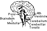
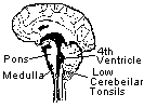
Arnold - Chiari, sometimes referred to as the Chiari Malformation is an anomaly of the brain, which occurs in almost all children born with spina bifida and hydrocephalus. It involves the lower brainstem and lowermost portion of the cerebellum.
The brainstem serves as the origin for many of the cranial nerves, which supply fibres to the head, eyes, neck and visceral organs. It is also the centre of control for the heart, breathing, blood pressure, as well as for swallowing, vomiting, sneezing and coughing. The cerebellum functions in the maintenance of posture and coordination of muscle action, to produce precise, coordinated movements. When the Chiari Malformation is present, the brainstem is elongated, and pushed down through the opening of the base of the skull. The brainstem, cranial nerves and the lower portion of the cerebellum may be stretched or compressed. Therefore, any of the functions controlled by these areas may be affected. Many children with Arnold - Chiari may present no obvious symptoms. In infants, the most common symptoms are stridor (a harsh croupy noise associated with breathing) and swallowing difficulties. Older children may present, but uncommonly, with upper limb weakness, and breathing difficulties. |
||||||
|
||||||
|
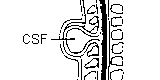
The normal spinal cord moves freely in the spinal canal. However, in spina bifida, commonly the spinal cord is tethered or "stuck down" at the site of the lesion, most often by scar tissue. This can cause stretching of the cord as the child grows.
POSSIBLE SYMPTOMS INCLUDE
All of these symptoms can be from other causes, and need to be investigated. Tethering is usually not significant unless a person presents with clinical symptoms. In some cases, it may be necessary to have an operation to "detether" the spinal cord. |
||||||
| ENCEPHALOCELE | Local herniation of neural tissue through a defect in the skull. Often, this will involve only the brain coverings, and children will have few problems. Sometimes, the brain is also involved. Hydrocephalus may also occur. | |||||
| SCOLIOSIS | A lateral curvature of the spine, often in an S shape. | |||||
| KYPHOSIS | An exaggerated outward curvature of the spine. | |||||
| KYPHOSCOLIOSIS | A spinal deformity, in which kyphosis and scoliosis occur together. This is often how spinal deformities present in spina bifida. | |||||
| CAT SCAN | An x-ray machine which revolves around the body, and takes a picture of the organs inside the body. | |||||
| Magnetic Resonance Imagery - a scanner using magnetic energy to give a clear black and white picture of the brain, and spinal cord. Does not involve radiation |
| Facts About Spina Bifida and Hydrocephalus |

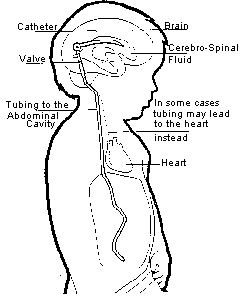 Hydrocephalus is usually treated by insertion of a "shunt". A shunt is a device which is designed to drain excess cerebrospinal fluid from the brain and carry it to other parts of the body. A one-way valve is used, which usually sits outside the skull, but beneath the skin, somewhere behind the ear. (see diagram)