The urinary system is one of the most vital in the human body. It has two functions: to filter waste and excess water from our blood to form urine and to return salt and other important chemicals to the blood. It is of extreme importance to your health that the urinary system function properly.
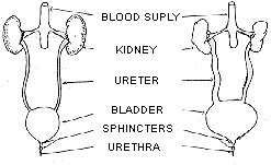
The urinary system consists of a pair of kidneys, two ureters, a bladder, a urinary sphincter muscle, and a urethra. The kidneys are made of special filtering tissue through which all of the body's blood passes several times a day. They strain the useful material from the blood and send excess water and waste material down two thin tubes (ureters) to the bladder. Each ureter tube has a flap of skin on the end of it which closes to prevent urine from flowing backwards up the ureters into the kidney.
| NORMAL EMPTYING OF BLADDER |
BLADDER EMPTYING INCOMPLETELY |
| Normal Ureters and Kidneys |
Dilated Ureters Enlarged Bladder Damaged Kidneys |
The bladder is a muscular lined sac that can hold up to three pints of urine. When the bladder becomes fully expanded and cannot store any more urine, sensory receptors alert the brain and the brain sends a message to the bladder muscles to contract. When the bladder contracts pressure builds up and urine is pushed out through a short tube, called the urethra, and is discharged from the body. The urethra is surrounded by a circular muscle, the urinary sphincter which tightens to hold the urine in, or relaxes, to allow urine to empty from the bladder and leave the body. This sphincter muscle is voluntary, allowing a person to choose when to urinate.
The neurogenic bladder can be either flaccid or spastic. A flaccid bladder is limp and cannot contract completely to force urine out. When the flaccid bladder becomes full, excess urine spills over and flows out of the body through the urethra. Urine dribbles out continually and when excess pressure is put on the bladder such as when laughing or crying this dribbling becomes more severe. However, the bladder never empties completely and some (residual) urine always remains.
Unlike the flaccid bladder, the spastic bladder does not store urine at all. The muscles that line this type of bladder are extremely sensitive and irritable. They contract and expel urine immediately after it enters the bladder. Even though the bladder is continually contracting, some urine almost always remains in the bladder.
The bladder is also influenced by the control of the urinary sphincter. If nerve damage exist, the sphincter muscle can be either too tight or too loose. When the sphincter muscle is tight, urine becomes trapped in the bladder and is often forced back up the ureters to the kidney. If the sphincter muscle is too loose, however, urine continually leaks out of the body.
The function of the bladder is also influenced by pressure. Pressure is needed to force urine out. Low pressure in the bladder will result in incomplete emptying; high pressure can force urine into the ureters and kidneys (reflux).
This backward flow of urine can be very damaging to the urinary system and especially the kidneys. Normally, a muscular flap on the ureters closes and once the urine flows out of the kidneys, it cannot flow back. However, the muscles that control this flap are often damaged and instead of following the path from the kidneys to the bladder and outside of the body, the urine flows back up the ureters to the kidney.
It is very important for a person's health that the bladder is completely emptied regularly. Urine that remains in the bladder provides an excellent breeding ground for bacteria which thrive on warm, damp conditions. Repeated, severe infections, over time, substantially damage the kidneys and impair their filtering capabilities.
The signs of a urinary tract infection are cloudy or discoloured urine, fever, chills and shakes, headache, fatigue, nausea, pain and an increased frequency and need to urinate. A person with spina bifida who has paralysis in the lower extremities should monitor the appearance of their urine carefully since they may not be able to feel the first warning signs - pain while urinating, for example - of a urinary tract infection. (Reference 6)
With spina bifida, urinary incontinence will usually affect those children who have the most involved type (myelomeningocele). It may also affect, though not usually, those children with the less involved types of spina bifida (occulta & meningocele).
- 1) Medication
- 2) Toilet timing /training program = REGULARITY
- 3) Adapted clothing & specific incontinence products e.g. pads, shields.
- 4) Possible operations e.g. bladder augmentation artificial sphincter perineal urethrostomy
- 5) Clean intermittent catheterisation *** Usually a combination of the above suggestions is required for successful urinary continence management.
The aim of clean intermittent catheterisation is to help manage the abnormal bladder function by improving bladder emptying, and reducing urinary leakage (wetting) and to assist in protecting the kidneys from damage.
The development of independent clean intermittent catheterisation skills is usually encouraged at kindy and preschool and formalised at primary school. It is generally accepted that most children learn self- catheterisation and are basically independent by 8 years of age (Grade 3).
It is usually expected that children will see appropriate doctors regularly, to monitor kidney and bladder function.
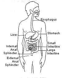
In a normally operating bowel the external sphincter will contract when the rectum is full and hold the faeces in the anal canal. However, because there is little or no control over the external anal sphincter for a person with spina bifida, faeces are often forced out of the body at an inappropriate time.
Limited sensation affects the ability to realize when the rectum is full. Nerve damage prevents the brain from receiving the message to empty the bowels. If it is not realized that the bowel is full and there is no control of the external sphincter then the bowels may open when it is least expected.

Being constipated can make one feel nauseated and grouchy and generally, very sick. Constipation occurs when the stool is unable to be removed from the body and becomes hardened because the water it contains is absorbed back into the body. (Reference 7)
- 1) Diet - high in fibre, plenty of water
- 2) Medication
- 3) Toilet timing, training program = REGULARITY
- 4) Enemas e.g. bowel washout, Microlax
- 5) Manual Evacuation
- 6) Exercise
- 7) Possible operations e.g. Mitrofanoff Antegrade Colonic Enema (M.A.C.E.)
The overall aims of bowel management programmes include:
- To reduce incontinence by establishing regular bowel evacuation
- To reduce the risk of a mega colon developing
- For the child to achieve "social continence" and be independent and confident in their bowel management strategies
- 1) Suitable toilet facilities: - wheelchair accessible (if necessary) - sink, cupboard and bin in toilet - ensure cleanliness of toilet area - ensure privacy
- 2) Aide time for supervision and/or assistance with catheterisation.
- 3) Extra time for toileting.
- 4) An established toilet routine.
- 5) To deal with accidents - change clothes - clean wheelchair - wash out soiled clothes
- 6) To be told they have had an accident as children are not always aware of the situation.
- 7) To inform the teacher if there is a change of management and the consequences.
- 8) To drink regularly to minimise urinary tract infections.
The system that is in place should always maintain the dignity of the child.
